This sections aims to provide a brief look at the invention of the microscope, and the resulting discoveries. Includes sections on the discovery of the cell. Also includes links to biographies of key persons who aided in discovery.
Cell History and Cell Theory
A PowerPoint lecture that I created on the basic discovery of cells and the cell theory. Also contains some information on early microscopes. Created from Pearson Education: Biology textbook, but is general enough to work with any textbook. There is also a compatible worksheet that deals with the Microscope and Early Discoveries (page 1) that is created and (c) Pearson Education.
If you take things a bit further in this section, and start to examine characteristics of living things, this computer project is a nice activity. It is also really simple, which allows for additions to be made to challenge students that need a little more. Students use the internet to search out images they can use as examples of each of the eight characteristics of living things and either create a poster, or you can have them put together a PowerPoint, or even use a movie program like Windows Movie Maker to create a video presentation.
If you take things a bit further in this section, and start to examine characteristics of living things, this computer project is a nice activity. It is also really simple, which allows for additions to be made to challenge students that need a little more. Students use the internet to search out images they can use as examples of each of the eight characteristics of living things and either create a poster, or you can have them put together a PowerPoint, or even use a movie program like Windows Movie Maker to create a video presentation.
Timeline of Discovery
A timeline of major events in microscopes and cell theory. Molecular Expressions is a really good website put together by Florida State Univ. It is easily a good site to recommend for student research on discovering in various time periods. Time Periods include:
Biographies
Founding Fathers of Microscopy:
Zacharias Janssen is accredited as the man who produced the first compound microscope. This is just one of the many biographical options available on the web. Another good biography on the web can be found at the History of the Microscope.
It is from Robert Hooke that we get the term "cell". when he looked at corn through a microscope, he saw what looked like tiny monk cells in a monastery, and described them as such. Again, this is just one of the many biographical options available. Another is also available on the History/Microscope site.
Anton van Leeuwenhoek was the first person to see "living cells" via a microscope. He looked at teeth scraping, and saw tiny living organisms that he termed "animalcules". We knew them today as bacteria. Again, just one option for biographical data. Another can be found here.
For a brief biography of each of the three founding fathers of microscopy, check out this page offered by Miami Univ. It provides one paragraph biographies. A great resource for the more internet challenged students, or for those that need things kept simple and offered on only one page rather than having to search different pages for it.
If students are interested in other significant contributors to the field of microscopy, they can check out this list from Miami Univ. or check out the timelines pages linked above.
Founding Fathers of Cell Theory:
Schleiden, Schwann and Virchow each contributes to our understanding of the cell theory. Schleiden was the discover that plants are made up of cells. Schwann was the first to discover the same for animals. From Virchow we get the cell theory - that all living things are made up of cells. A brief biography of all three can be found here - offered by Brooklyn College in NY.
Handouts and Lab Activities
Cell Theory: A handout on Schleiden and Schwann's contribution to the cell theory. This is a really good handout for students that aren't comfortable with using the internet for research, or for special education students that need to have hard copies of articles to read.
Early Experiments in Germ Theory
Spontaneous Generation was disproved primarily by the work of three scientists - Redi, Spallanzani, and Pasteur. I have included information on each of their contribution. Some are graphics and some are videos or tutorials from various sources.
Redi - His experiment involved covered and uncovered jars with meat in them. In the uncovered jars, maggots appears after a few days - much like had been seen over the years. In the covered jars however, no maggots appeared. What I like about the first image is that it breaks down the "scientific method" for Redi's experiment - showing not only what was done, but explaining each step of the scientific method that occurred The second image for Redi's experiment focuses just on the control v experimental/treatment groups of the experiment.
Redi - His experiment involved covered and uncovered jars with meat in them. In the uncovered jars, maggots appears after a few days - much like had been seen over the years. In the covered jars however, no maggots appeared. What I like about the first image is that it breaks down the "scientific method" for Redi's experiment - showing not only what was done, but explaining each step of the scientific method that occurred The second image for Redi's experiment focuses just on the control v experimental/treatment groups of the experiment.
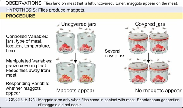
Image 1: Redi's Experiment to disprove spontaneous generation
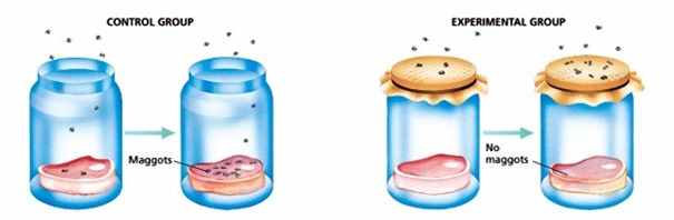
Image 2: Focusing on the control v treatment/experimental groups of Redi's experiment.
Spallanzani boiled gravy broth and then left one flash open and sealed another. The boiling process killed off anything that might have already been living int he broth. In the open flask, microorganisms appeared, but in the sealed flask they did not. If Spontaneous Generation were correct, they should have. Image 3 is just a real simple explanation of Spallanzani's experiment.
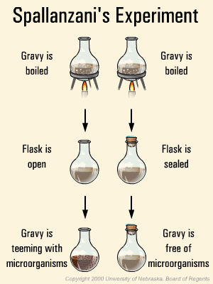
Image 3: Spallanzani's experiment.
Pasteur's famous experiment involved taking Spallanzani's one step further, using a swan-neck flask. There is a really nice informational tutorial offered here. It includes a brief introduction, and then the choice of a self-guided or narrated animation. At the end is a brief "quiz" to test for understanding. I have also included a couple of images of his experiment that can be found on the internet. Each of them has something to recommend it. Image 4 is very simple and easy to understand - with the curved neck, no microorganisms could get into the flask, but once it was broken, they could. Image 5 offers a bit more, with the two hypotheses being examined - Germ Theory and Spontaneous Generation.

Image 4: Pasteur's experiment simplified.
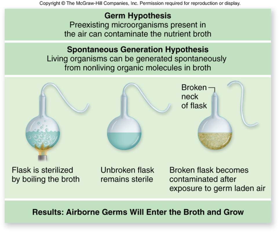
Image 5: Pasteur's experiment with the two different hypotheses - Germ Theory and Spontaneous Generation.
